An upper GI endoscopy is not a routine test for finding an incisional hernia. You will learn when symptoms or medical history justify it, which test is better for examining the abdominal-wall defect, how to prepare safely, and how results could affect surgery timing and anaesthesia planning.
Key takeaways
- Endoscopy examines the oesophagus, stomach and duodenum—not the abdominal-wall hernia.
- Hernia assessment uses examination, ultrasound or CT when imaging is needed.
- Ask why endoscopy is recommended and whether its findings could alter surgery.
- Fast as instructed and bring scans, reports, medicines and previous operation records.
Is an Upper GI Endoscopy Necessary Before Incisional Hernia Surgery?
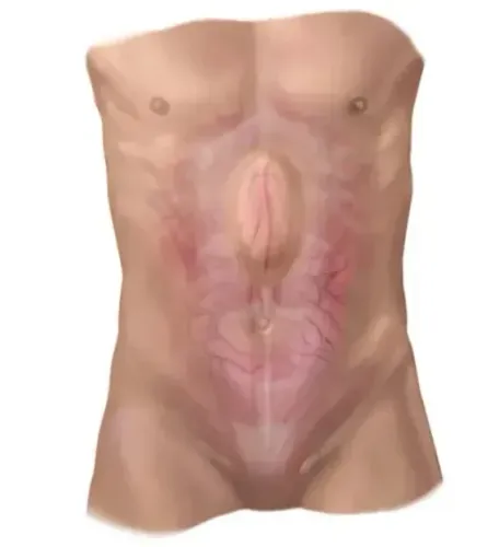
Upper GI endoscopy is not routinely required just because incisional hernia repair is planned. Searching upper GI endoscopy before incisional hernia for surgery in Nagpur should not imply that endoscopy diagnoses the hernia: it examines the oesophagus, stomach and duodenum, while the hernia is assessed by abdominal-wall examination and, when needed, ultrasound or CT.
Arrange endoscopy when you have a separate upper-digestive concern, such as:
- Difficulty swallowing
- Gastrointestinal bleeding or iron-deficiency anaemia
- Unexplained weight loss
- Persistent vomiting
- Significant dyspepsia
- Suspected ulcer disease
- Reflux complications
Do not wait for elective endoscopy if an incisional hernia becomes an emergency. An irreducible painful lump, rapidly increasing swelling, vomiting, abdominal distension, fever, or inability to pass stool or gas requires urgent assessment because trapped or strangulated bowel is possible.
A normal endoscopy does not prove that the hernia is safe to repair and does not exclude bowel involvement. It only shows that no relevant abnormality was seen in the upper digestive tract. Conversely, a significant abdominal-wall hernia can exist with a completely normal oesophagus, stomach and duodenum.
Which Test Examines the Hernia and Which Examines the Upper Digestive Tract?
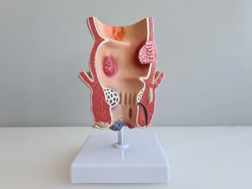
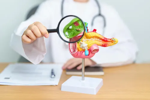
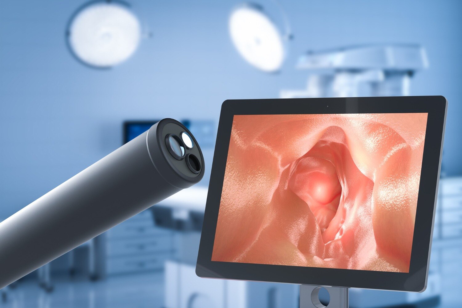
An upper GI endoscopy examines the upper digestive tract, not the abdominal-wall hernia. A flexible gastroscope passes through your mouth to view the oesophagus, stomach and duodenum; it does not enter the old abdominal incision, diagnose the fascial defect, or repair the hernia.
During examination, the surgeon checks the lump’s position and size, whether it reduces when you lie down or with gentle pressure, tenderness, the old operation site, previous mesh or infection, and bowel symptoms such as vomiting, constipation or difficulty passing gas.
This clinical assessment often provides the starting point for upper GI endoscopy incisional hernia planning.
| Test | What it examines | When it helps |
|---|---|---|
| Upper GI endoscopy | Oesophagus, stomach and duodenum | Upper-digestive symptoms or suspected disease |
| Abdominal examination | Lump, reducibility, tenderness and incision | Every incisional-hernia assessment |
| Ultrasound | Abdominal-wall movement and a fascial gap | Smaller or uncertain defects |
| CT scan | Defect, contents, loss of domain, muscle quality and other abdominal abnormalities | Large, recurrent, complex or obese-patient hernias |
CT gives the surgeon a map for planning but does not automatically mean surgery is required. The same distinction applies to hernia testing in Nagpur: ultrasound or CT assesses the abdominal wall, while endoscopy answers a separate digestive-tract question.
What Can Endoscopy Find, and When Can It Change the Operation Plan?
An upper GI endoscopy before incisional hernia for surgery in Nagpur can uncover an upper-digestive problem that deserves treatment before elective repair. It does not assess the abdominal-wall defect itself.
| Finding | Possible effect on the plan |
|---|---|
| Reflux damage or severe oesophagitis | Change acid-treatment instructions, aspiration precautions or timing |
| Gastritis or peptic ulcer disease | Add treatment; active ulcer bleeding can delay repair |
| Bleeding source | Treat or investigate before anaesthesia |
| Narrowing or gastric outlet obstruction | Require further treatment and postpone hernia surgery |
| Suspected hiatal hernia | Guide symptom treatment and anaesthetic planning |
| Suspicious lesion | Require biopsy, pathology and a separate treatment decision |
| Normal examination | Clarify upper-digestive symptoms but does not determine the hernia repair |
A biopsy does not itself diagnose cancer. A pathologist must examine the tissue, and the result can change the next step.
Severe oesophagitis, active bleeding, obstruction, a suspicious lesion or another untreated condition can delay hernia repair. The findings can also change medicines, fasting instructions, aspiration precautions, anaesthetic planning or the need for further tests.
Separate optimisation issues can delay elective surgery even when endoscopy is normal. Smoking raises wound and pulmonary risk; poor diabetes control increases infection and impaired healing; obesity can increase wound problems and recurrence; malnutrition weakens healing; respiratory disease raises the risk of postoperative breathing complications. These factors are assessed independently of endoscopy.
How to Prepare for the Endoscopy and What the Procedure Feels Like
Follow the endoscopy unit’s written instructions rather than a universal timetable. Many units request at least 6 hours without solid food and allow clear liquids until 2 hours before sedation or anaesthesia. Diabetes, delayed stomach emptying and the planned procedure can change these times.
Ask specifically how to take insulin, diabetes tablets, reflux medicines and blood-pressure medicines. Do not stop warfarin, apixaban, rivaroxaban or clopidogrel independently. The endoscopist and prescribing clinician decide the plan according to your clotting risk and whether biopsy or treatment is planned.
- At arrival, confirm your medicines, allergies and medical conditions, then discuss the procedure and sign consent. Ask about throat spray, intravenous sedation and possible biopsy.
- Throat spray numbs the back of your mouth. With sedation, you become relaxed and drowsy; without it, you remain awake.
- The endoscopist passes a thin flexible gastroscope through your mouth to examine the oesophagus, stomach and duodenum. The examination is brief. Gagging and abdominal pressure are common, but the scope does not block normal breathing.
- Afterward, rest while staff check you. A sore throat can last for several hours.
Arrange an escort if you receive sedation, even when the upper GI endoscopy is brief. For the rest of that day, do not drive, operate machinery, sign important documents or perform safety-critical work. These precautions concern the endoscopy; they do not assess or repair an incisional hernia.
What to Take to an Incisional Hernia Surgery Consultation in Nagpur
Bring the complete record, not only the latest scan. For an incisional hernia surgery consultation, organise:
- Previous operation report and discharge papers, including the incision location and date.
- Mesh brand or placement details, wound infection records and treatment notes.
- CT or ultrasound images and written reports.
- Endoscopy reports, biopsy results and pathology reports.
- Every medicine, dose and timing, including warfarin, apixaban, rivaroxaban, clopidogrel, insulin and diabetes tablets.
- Drug, food and latex allergies, smoking history, diabetes information and recent glucose results.
- A symptom record covering vomiting, bleeding, difficulty swallowing, abdominal distension, constipation, inability to pass gas and other bowel changes.
These documents make hernia testing in Nagpur more efficient. The surgeon can decide whether examination alone is enough, whether ultrasound or CT is needed, whether an endoscopy finding requires treatment first, and whether you need blood tests, an ECG, anaesthesia review or chest assessment based on your age, illnesses and planned operation.
| Information available | Decision it supports |
|---|---|
| Examination and operation history | Whether the defect is reducible and how repair may be planned |
| CT or ultrasound | Defect size, contents and complexity |
| Endoscopy and pathology | Whether upper-digestive treatment comes before surgery |
A consultation with Dr Hemant Chhajed Surgeon can connect upper-digestive findings with abdominal-wall assessment rather than treating endoscopy as hernia evaluation. Ask who will coordinate medicine changes. Before the appointment, seek immediate care for a painful irreducible lump, repeated vomiting, fever, increasing swelling, abdominal distension, or inability to pass stool or gas.
Frequently asked questions
Is upper GI endoscopy necessary before incisional hernia surgery?
No. It is not routinely required for every incisional hernia repair. A surgeon recommends it when upper digestive symptoms, anaemia, bleeding or another clinical concern needs investigation.
Which test examines an incisional hernia?
A physical abdominal-wall examination assesses the hernia. Ultrasound or CT can show the defect, its contents and the surrounding abdominal wall when examination is insufficient.
What does an upper GI endoscopy examine?
A flexible camera examines the oesophagus, stomach and duodenum. It does not diagnose the abdominal-wall defect itself.
How should you prepare for an upper GI endoscopy?
Follow the clinic’s fasting instructions, disclose every medicine and allergy, and arrange transport if sedation is planned. Bring previous reports and operation records.
What should you take to an incisional hernia surgery consultation in Nagpur?
Take CT or ultrasound images and reports, previous surgical notes, discharge summaries, current medicine details, allergy information and a list of symptoms.
Keywords